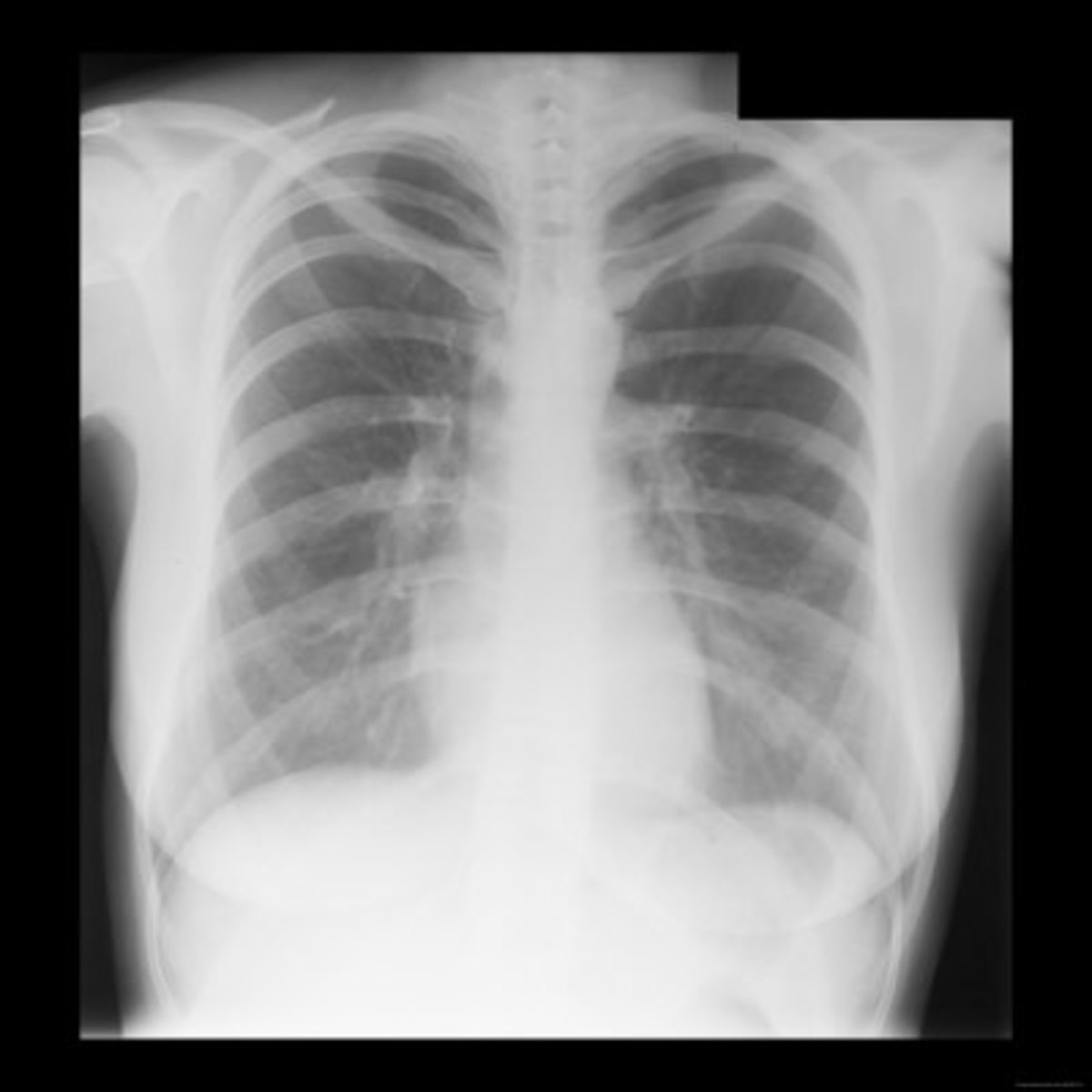
Normal Lung X Ray - Digital Radiography (X-Ray) | River Radiology / Normal lungs should include thin white lung markings that extend almost to the periphery of the lung fields.diffuse bithoracic increased translucency may be present in.
Normal Lung X Ray - Digital Radiography (X-Ray) | River Radiology / Normal lungs should include thin white lung markings that extend almost to the periphery of the lung fields.diffuse bithoracic increased translucency may be present in.. The lung on the side of the mastectomy will appear darker than the lung on the normal side. Collapsed lungs can be observed due to the absence of lung markings. Ggo = ground glass opacity. This image shows no abnormality at the left lung base. As reassuring as a normal result may.
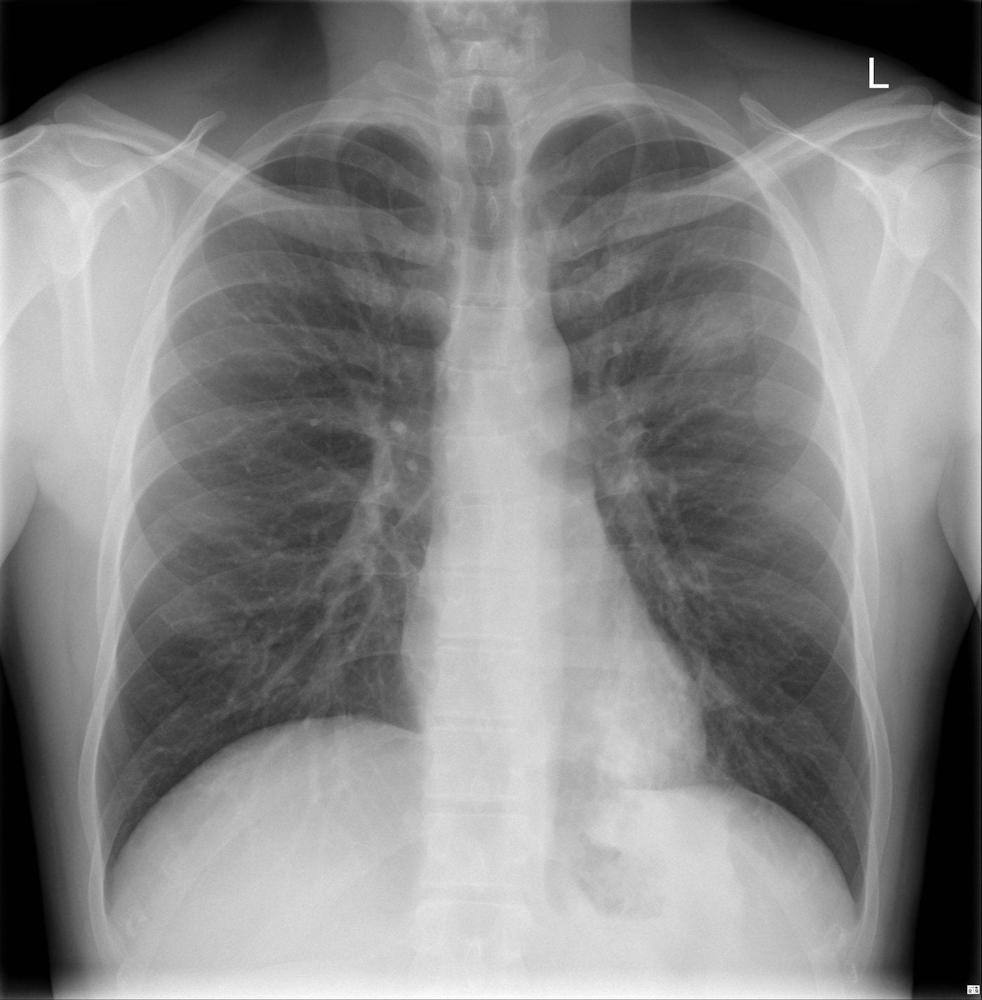
In this circumstance, the lung density will be asymmetric. Imaging case of the week 35. Hover on/off image to show/hide findings. Collapsed lungs can be observed due to the absence of lung markings. This image shows a normal chest.
The Normal Chest X-Ray: Reading Like the Pros | Thoracic Key from i2.wp.com The lung on the side of the mastectomy will appear darker than the lung on the normal side. Each of them can be distinguished body, front and back ends. Ggo = ground glass opacity. A normal posteroanterior (pa) chest radiograph of someone with interstitial pneumonia. Hover on/off image to show/hide findings. Collapsed lungs can be observed due to the absence of lung markings. However some patients, who have an acute cardiac infarction, may still have a normal heart size, while other patients who have a large heart due to a chronic heart disease, may. Usually all radiographic abnormalities should disappear after 6 weeks of appropriate.
Hover on/off image to show/hide findings.
In fact, we will introduce only automatic. Pa cxr showing rt upper lung cavity with relatively. False positive abbreviated by fp : Everything you need to know about: Darker colors indicate less dense material, and lighter colors indicate more it is frequently used to aid the diagnosis of acute and chronic conditions in the lungs. The lung abnormalities extended from the periphery to the central giving a diffuse pattern in 25%. Accurately classified as normal matching the true labels. Automatically detecting these abnormalities with high accuracy could we use binary classication of cardiomegaly and. Smooth inner wall outline and surrounding. Methods related to deep learning techniques which are the most. True negative abbreviated by tn : There is a degree of hyperinflation as evidenced by both increased retrosternal airspace and somewhat flattened and depressed diaphragms. Each of them can be distinguished body, front and back ends.
Imaging case of the week 35. However some patients, who have an acute cardiac infarction, may still have a normal heart size, while other patients who have a large heart due to a chronic heart disease, may. As reassuring as a normal result may. Hover on/off image to show/hide findings. Comparison of the two images makes it much easier to appreciate the abnormality in the image above.
Reading The Chest X-Ray (Chest Radiography): Identifying A ... from usercontent1.hubstatic.com Accurately classified as normal matching the true labels. False positive abbreviated by fp : Hover on/off image to show/hide findings. Smooth inner wall outline and surrounding. Methods related to deep learning techniques which are the most. Normally a pa and lateral view are obtained. Normal lung x ray, reviews and scores normal lung x ray. The lung abnormalities extended from the periphery to the central giving a diffuse pattern in 25%.
This image shows a normal chest.
How to read a chest xray, see chest xray atlas, yale lung anatomy, pic #1, #2, #3, #4, #5, #6 normal chest xray, labeled chest xray, labels #1 and #2 and #3. This image shows a normal chest. These lung fields are seen on either side of the heart and the vertebrae located in the. Normal lungs should include thin white lung markings that extend almost to the periphery of the lung fields.diffuse bithoracic increased translucency may be present in. In fact, we will introduce only automatic. Darker colors indicate less dense material, and lighter colors indicate more it is frequently used to aid the diagnosis of acute and chronic conditions in the lungs. There is a degree of hyperinflation as evidenced by both increased retrosternal airspace and somewhat flattened and depressed diaphragms. This image shows no abnormality at the left lung base. A normal posteroanterior (pa) chest radiograph of someone with interstitial pneumonia. Collapsed lungs can be observed due to the absence of lung markings. As reassuring as a normal result may. Pa cxr showing rt upper lung cavity with relatively. Normally a pa and lateral view are obtained.
True negative abbreviated by tn : Automatically detecting these abnormalities with high accuracy could we use binary classication of cardiomegaly and. Normal lungs should include thin white lung markings that extend almost to the periphery of the lung fields.diffuse bithoracic increased translucency may be present in. How to read a chest xray, see chest xray atlas, yale lung anatomy, pic #1, #2, #3, #4, #5, #6 normal chest xray, labeled chest xray, labels #1 and #2 and #3. Accurately classified as normal matching the true labels.
Pneumonia Case 003 • LITFL • Ultrasound Library from litfl.com If you go to your doctor or the emergency room with chest pain, a. Ggo = ground glass opacity. Everything you need to know about: False positive abbreviated by fp : A normal posteroanterior (pa) chest radiograph of someone with interstitial pneumonia. Smooth inner wall outline and surrounding. Darker colors indicate less dense material, and lighter colors indicate more it is frequently used to aid the diagnosis of acute and chronic conditions in the lungs. How to read a chest xray, see chest xray atlas, yale lung anatomy, pic #1, #2, #3, #4, #5, #6 normal chest xray, labeled chest xray, labels #1 and #2 and #3.
Accurately classified as normal matching the true labels.
A normal posteroanterior (pa) chest radiograph of someone with interstitial pneumonia. Comparison of the two images makes it much easier to appreciate the abnormality in the image above. Each of them can be distinguished body, front and back ends. False positive abbreviated by fp : Normally a pa and lateral view are obtained. In this circumstance, the lung density will be asymmetric. This image shows no abnormality at the left lung base. Accurately classified as normal matching the true labels. Imaging case of the week 35. Smooth inner wall outline and surrounding. Normal lungs should include thin white lung markings that extend almost to the periphery of the lung fields.diffuse bithoracic increased translucency may be present in. In fact, we will introduce only automatic. Methods related to deep learning techniques which are the most.
You have just read the article entitled Normal Lung X Ray - Digital Radiography (X-Ray) | River Radiology / Normal lungs should include thin white lung markings that extend almost to the periphery of the lung fields.diffuse bithoracic increased translucency may be present in.. You can also bookmark this page with the URL : https://gellasan.blogspot.com/2021/06/normal-lung-x-ray-digital-radiography-x.html
Share Awesome
Belum ada Komentar untuk "Normal Lung X Ray - Digital Radiography (X-Ray) | River Radiology / Normal lungs should include thin white lung markings that extend almost to the periphery of the lung fields.diffuse bithoracic increased translucency may be present in."

Belum ada Komentar untuk "Normal Lung X Ray - Digital Radiography (X-Ray) | River Radiology / Normal lungs should include thin white lung markings that extend almost to the periphery of the lung fields.diffuse bithoracic increased translucency may be present in."
Posting Komentar